I.V. House Products Improve Site Access to Prevent Grades 3 and 4 Infiltrates
With increasing strain on hospitals, and health care professionals leaving the industry, we know that nurses are busier than ever. Best-practice guidelines say pediatric and critically ill adult patients receiving IV therapy require the IV insertion site be thoroughly assessed every hour to check for warning signs of complications.
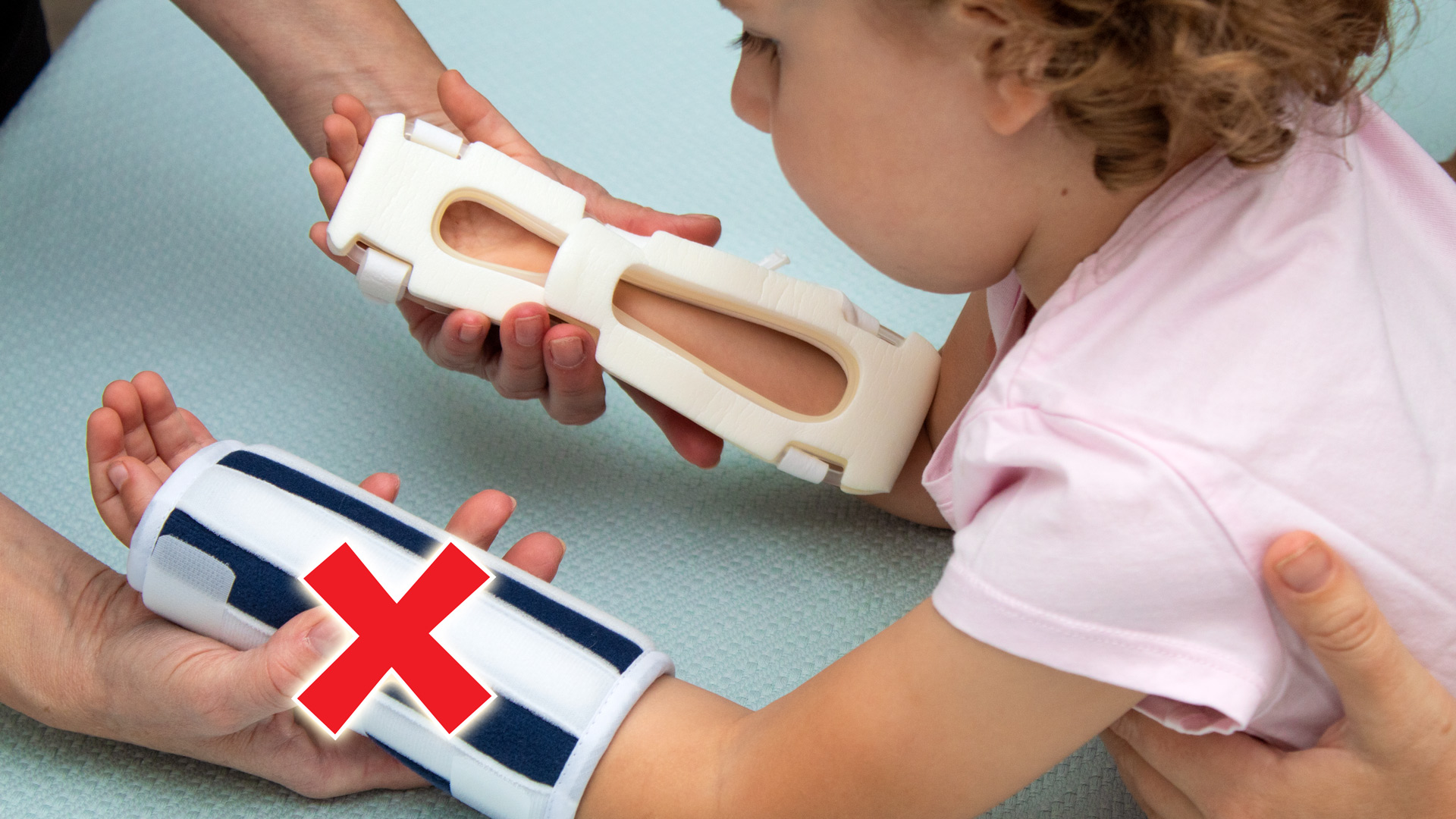
Traditional armboards and other immobilizers require removal in order to get a 360-degree view of the extremity with the IV catheter—a time-consuming process. Removing these devices for routine assessments can cause patients to wake unnecessarily during their hospitalization. In addition, immobilizers that wrap around the extremity and attach with hook and loop fasteners can be wrapped too tightly, causing pressure injuries. Nurses need faster access to the IV insertion site to be able to effectively spot the early warning signs of complications.
The Importance of Assessment
Studies show routine assessment of peripheral catheter insertion sites is the most effective process improvement in the early detection of IV infiltration.1 In a recent study, a neonatal intensive care unit experienced two stages 3 to 4 infiltrates in two months. Both cases were with pre-term infants that were unable to communicate their level of discomfort. The hospital protocol stated that a clinically stable infant may have the intravenous site assessed every four hours. In both cases, documentation of site assessments was either infrequent or exceeded four hours.2
Lack of documentation is a serious concern. Results from an IV catheter monitoring questionnaire showed that the most prevalent overall problem with IV therapy is lack of documentation of the PIVC, prompting the response, “In nursing school we are taught if you didn’t document, it wasn’t done.”3
The Role of Patients and Caregivers
Patients and their parents/caregivers rely on nursing staff to monitor PIVCs for warning signs of complications, however, health care organizations find incorporating these parties into the health care team helps reduce patient harm. Frequent assessment of peripheral IV sites by patients and their family members can help detect infiltration/extravasation in the early stages. A 2015 study showed significantly lower incidence of pediatric IV infiltrations when guardians were educated about the warning signs associated with common complications.4 Utilizing a mnemonic device, such as a memorable word or phrase, along with educational signage empowers patients and their caregivers to speak up if something seems wrong.
The awareness of the problem is worldwide, and although evidence-based trials are still needed, hospitals that switch from opaque immobilizers and traditional armboards to I.V. House products report fewer grades 3 to 4 infiltrates.
Preventing Grades 3 and 4 Infiltrates
Because I.V. House products are designed specifically for IV therapy to help improve patient safety and increase nurse efficiency, hospitals that convert from traditional armboards and immobilizers to the TLC Splint see a reduction in grades 3 and 4 infiltrates.
The TLC® Splint, available for the wrist, elbow, and foot, has see-through openings that allow nurses to Touch, Look, and Compare the IV insertion site with the opposite extremity without removing the device, making visual and manual assessment much faster. Nurses, patients, and their family members can check for early warning signs of complications such as changes in color, temperature, or swelling by visualizing the site, and palpating the surrounding tissue.
Protecting the IV Catheter from Dislodgment
Peripheral IV catheter (PIVC) loss is common and millions of patients are affected each year. In addition to joint stabilization, IV catheter securement can minimize PIVC movement within the vessel, preventing irritation, occlusion, infiltration, infection, or dislodgment that culminate in PIVC failure. Using an IV catheter securement device that prevents the PIVC from falling out in partnership with a joint stabilization device reduces the chance for accidental dislodgment and early PIVC failure.5
The I.V. House UltraDome® and I.V. House UltraDressing® IV insertion site protection products offer clear, ventilated plastic domes that can be easily lifted to see the IV catheter and surrounding tissue while protecting the site from bumps and snags that can lead to accidental dislodgment.
Conclusion
When a vesicant is extravasated it can inflict severe and permanent tissue damage ranging from skin blanching, swelling, pain, blisters, vascular occlusion, phlebitis, and if allowed to progress can lead to frank necrosis, gangrene, and compartment syndrome.6 Nurse training, patient and caregiver education, and improved visibility of the venous access sites allow nurses to spot the signs of potential complications before they escalate. The awareness of problems associated with IV therapy is worldwide, and although evidence-based trials are still needed, hospitals that switch from opaque immobilizers and traditional armboards to I.V. House products report fewer grades 3 and 4 infiltrates.
Please contact us for samples or to set up a trial of I.V. House products.
- The Effect of Intravenous Infiltration Management Program for Hospitalized Children. (2015). Park, SM, PhD, RN, et al. Journal of Pediatric Nursing, Vol 31, Issue 2, P. 172-178
- Quality improvement measures for early detection of severe intravenous infiltration in infants. (2019). Sangam, SL BMJ Open Quality Improvement Report 2019;8:e000407
- Monitoring quality of care for peripheral intravenous catheters; feasibility and reliability of the peripheral intravenous catheters mini questionnaire (PIVC-miniQ). (2019). Høvik et al. BMC Health Services Research
- S.T.I.C.K.: A Quality Improvement Pediatric IV Infiltration Prevention Bundle. (2018). Watterson, K, BSN RN, CPN, et al. Journal of Pediatric Nursing, Vol 41, P. 38-41
- Peripheral intravenous catheter duration and failure in paediatric acute care: A prospective cohort study. (2014). Malyon, L. et al. Emergency Medicine Australasia
- Extravasation Injuries: A Trivial Injury Often Overlooked with Disastrous Consequences. (2020). Alexander, L M.D, World Journal of Plastic Surgery, Vol. 9, No 3.